Anatomical subendocardical fibers distribution in mammals, first report
Abstract
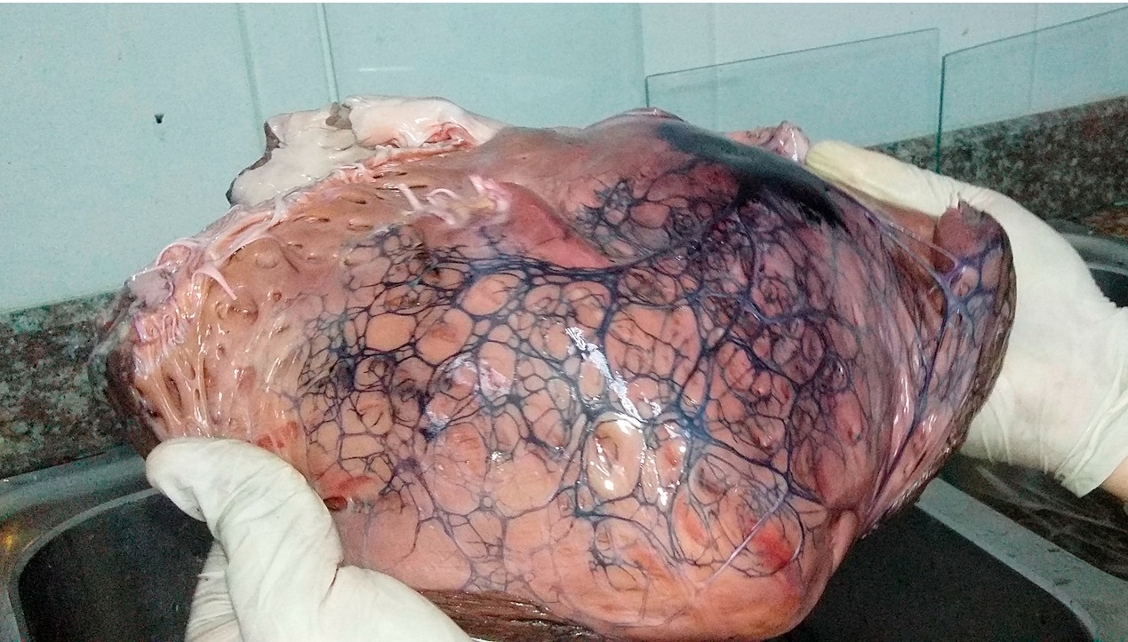
Establecer la organización de las fibras subendocárdicas es útil para determinar la conducción y propagación normal o anormal del impulso cardiaco y tiene trascendencia en el campo educativo, hemodinámico y cardiológico; su identificación ha sido posible habiéndose encontrado patrones similares en muchas especies mamíferas. Históricamente denominadas fibras de Purkinje, las ramas subendocárdicas (A.12.1.06.008) de Terminologia Anatomica son inidentificadas macroscópicamente y han sido evidenciadas con varias técnicas con distinto valor y limitaciones. Como componente de la formación académica, en el laboratorio de Anatomía Humana de la Universidad de Guayaquil se ha replicado su identificación utilizando tinta china en corazones adultos de vacunos y porcinos frescos mostrándose macroscópica e histológicamente su presencia. El nodo sinoatrial y atrioventricular son uniformes en su constitución, las fibras subendocárdicas muestran una estructura reticular con múltiples interconexiones, esto ha permitido realizar una descripción aproximada. Sus detalles de distribución y organización son importantes para interpretar los problemas de conducción como las arritmias.
Downloads
References
-Tawara, S., SHIMADA, M. & SUMA, K. (. The conduction system of the mammalian heart: an anatomico-histological study of the atrioventricular bundle and the Purkinje fibers. Imperial College Press,. 2000.
- Murillo, M., Cabrera, J. A., Pizzaro, G. & Sánchez-Quintana, D. Anatomía del tejido especializado de conducción cardíaco. Su interés en la cardiología intervencionista. RIA, 1(2), 229-245. 2011.
-Truex, R. C., & Smythe, M. Q. Comparative morphology of the cardiac conduction tissue in animals. Annals of the New York Academy of Sciences, 127(1), 19-33. 1965.
- Ono, N., Yamaguchi, T., Ishikawa, H., Arakawa, M., Takahashi, N., Saikawa, T., & Shimada, T. Morphological varieties of the Purkinje fiber network in mammalian hearts, as revealed by light and electron microscopy. Archives of histology and cytology, 72(3), 139-149. 2009.
-Waller, B. F., Gering, L. E., Branyas, N. A. & Slack, J. D. (). Anatomy, histology, and pathology of the cardiac conduction system: Part II. Clinical cardiology, 16(4), 347-352. 1993.
-Widran, J.& Lev, M. The dissection of the atrioventricular node bundle and bundle branches in the human heart. Circulation, 4(6), 863-867. 1951.
-Ansari, A., Yen Ho, S., & Anderson, R. H. Distribution of the Purkinje fibres in the sheep heart. The Anatomical Record: An Official Publication of the American Association of Anatomists, 254(1), 92-97. 1999.
-De Almeida, M. C., Lopes, F., Fontes, P., Barra, F., Guimaraes, R. & Vilhena, V.. Ungulates heart model: a study of the Purkinje network using India ink injection, transparent specimens and computer tomography. Anatomical science international, 90(4), 240-250. 2015
- Montalvo, C.E. .Técnica histológica; http://bct.facmed.unam.mx/wp-content/uploads/2018/08/3_tecnica_histologica.pdf. 2010.
-Abramson, D. I., & Margolin, S. . A Purkinje conduction network in the myocardium of the mammalian ventricles. Journal of anatomy, 70(Pt 2), 250. 1936.
-De Almeida, M. C., Araujo, M., Duque, M. & Vilhena, V.. Crista supraventricularis Purkinje network and its relation to intraseptal Purkinje network. The Anatomical Record, 300(10), 1793-1801. 2017.
-Sedmera, D., & Gourdie, R. G. Why do we have Purkinje fibers deep in our heart?. Physiological research, 63. 2014.
-Cardwell, J. C., & Abramson, D. I. The atrioventricular conduction system of the beef heart. American Journal of Anatomy, 49(2), 167-192. 1931.
-Hondeghem, L. M. & Stroobandt, R. Purkinje fibers of sheep papillary muscle: occurrence of discontinuous fibers. American Journal of Anatomy, 141(2), 251-261. 1974.
-Uhley, H. N. & Rivkin, L. M. Visualization of the left branch of the human atrioventricula bundle. Circulation, 20(3), 419-421. 1959.
-Oosthoek, P. W., Viragh, S., Lamers, W. H. & Moorman, A. F. Immunohistochemical delineation of the conduction system. II: The atrioventricular node and Purkinje fibers. Circulation research, 73(3), 482-491. 1993.

Copyright (c) 2021 Rafael Coello Cuntó

This work is licensed under a Creative Commons Attribution 4.0 International License.
The authors retain the copyright, but assign to the Journal the rights of publication, edition, reproduction, distribution, exhibition and communication at the national and international level in the different databases, repositories and portals.


