
Revisión anatómica del cuero cabelludo
DOI:
https://doi.org/10.51343/revperuanamorfologia.v1i2.287Palabras clave:
cráneo, irrigación sanguínea, cuero cabelludo, neuralgia occipitalResumen
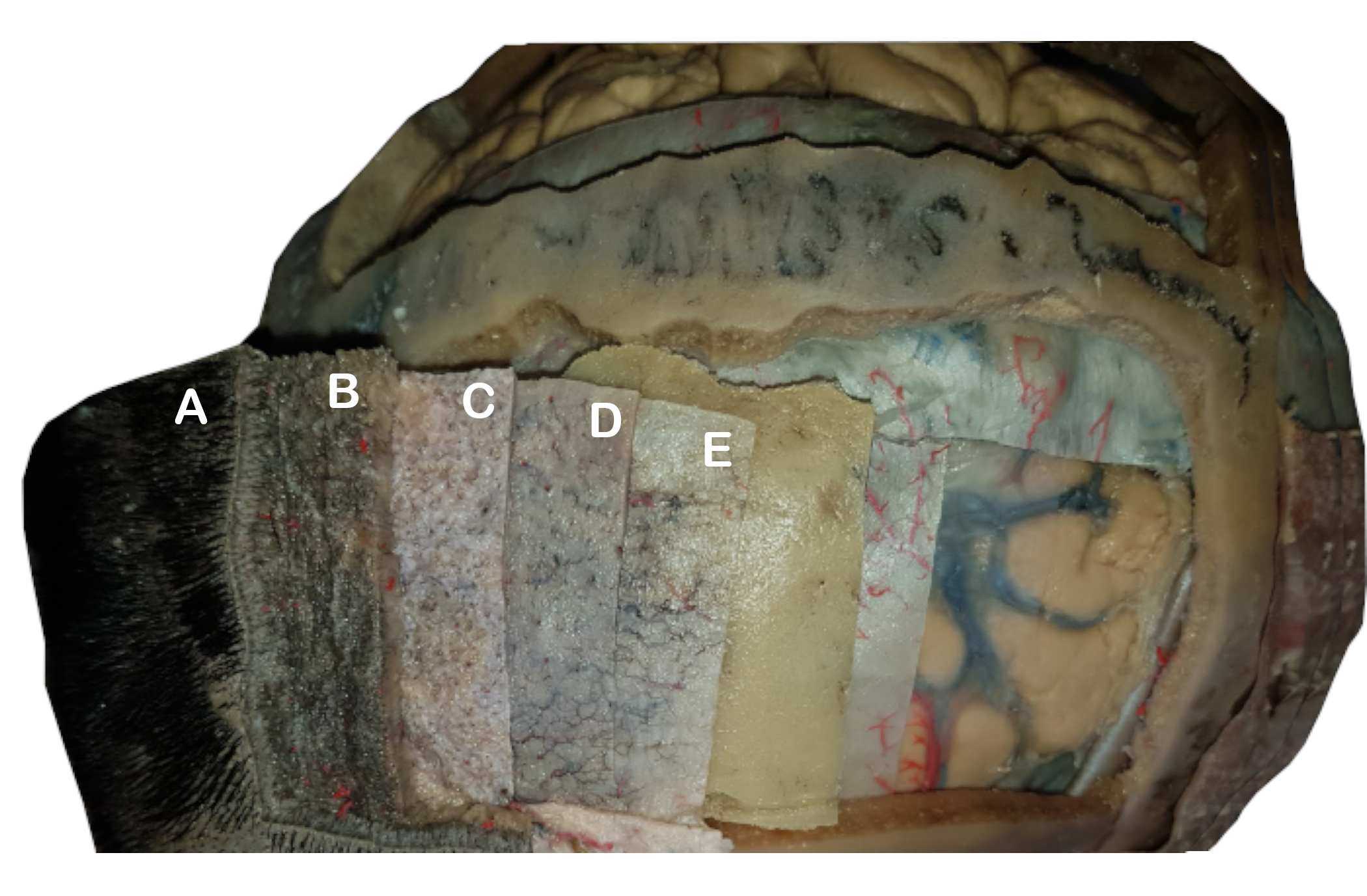
El cuero cabelludo es una estructura formada por cinco capas que rodean los huesos craneales, distribuidos desde la superficie encontramos las primeras tres estructuras unidas firmemente, estas son: piel, tejido conectivo denso y la gálea aponeurótica, la cual se separa del pericráneo por medio de tejido conectivo laxo. La inervación está suministrada por dos de los nervios craneales (facial y trigémino) y los nervios del plexo cervical. Se estableció un límite (orejas y vértice de la cabeza), el cual divide la inervación en una región anterior y posterior.
Esta región comprende de diversas arterias que se anastomosan entre sí formando un gran sistema que brinda un suministro sanguíneo característico; de modo parecido presenta un sistema de retorno venoso paralelo al arterial y acompañado del linfático.
Descargas
Citas
Iribarren O. Reconstrucción de cuero cabelludo. Cuad Cir. 2018;14(1):80–89.
Hayman AL, Shukla V, Ly C, Taber KH. Clinical and imaging anatomy of the scalp. J Comput Assist Tomogr. 2003;27(3):454–459.
Rukwied R. Physiology of the scalp. Hautarzt Z Dermatol Venerol Verwandte Geb. 2017;68(6):431–436.
Tajran J, Gosman A. Anatomía, cabeza y cuello, cuero cabelludo - Libros - NCBI [Internet]. [citado el 6 de junio de 2020]. Disponible en: https://www.ncbi.nlm.nih.gov/books/NBK551565/
Castañeda P, López S. El pelo: generalidades y enfermedades más comunes [Internet]. [citado el 6 de junio de 2020]. Disponible en: http://www.scielo.org.mx/scielo.php?script=sci_arttext&pid=S0026-17422018000300048&lng=es
Aldana C, Insfrán W, Sandoval J, Balmelli B, Aldana C, Insfrán W, et al. “RECONSTRUCCIÓN DEL CUERO CABELLUDO”-. Cir Paraguaya. agosto de 2018;42(2):25–7.
Gola R. [The forehead cutaneo-musculo-aponeurotic unit and aging of the forehead. Anatomo-physiological considerations and surgical implications]. Ann Chir Plast Esthet. febrero de 1999;44(1):89–102.
Balaguer-Cambra J, Landín Jarillo L, Hidalgo Gallego JC, Francés Gorospe MJ, Codina García J. Reconstrucción de cuero cabelludo mediante colgajo de galea frontal: a propósito de un caso. Cir Plástica Ibero-Latinoam [Internet]. marzo de 2006 [citado el 6 de junio de 2020];32(1). Disponible en: http://scielo.isciii.es/scielo.php?script=sci_arttext&pid=S0376-78922006000100008&lng=en&nrm=iso&tlng=en
Rouvière H, Delmas A, Delmas V. Anatomía humana: descriptiva, topográfica y funcional. Tomo 1. Décima Edición. Barcelona: Masson; 1999.
Navarro Cuellar C, Riba García F, Guerra Martínez B, Pujol Romanya R, Herencia Nieto H, Navvarro Vila C. Reconstrucción de cuero cabelludo con colgajo libre de omentum. Rev Esp Cir Oral Maxilofac. agosto de 2004;26(4):249–56.
Germann AM, Al Khalili Y. Anatomy, Head and Neck, Scalp Veins. En: StatPearls [Internet]. Treasure Island (FL): StatPearls Publishing; 2020 [citado el 3 de junio de 2020]. Disponible en: http://www.ncbi.nlm.nih.gov/books/NBK540961/
Gallardo J, Pessa D. Bloqueo del cuero cabelludo. Rev Chil Anest. 2013;7:42.
Drake RL, Vogl W, Mitchell AWM, Gray H. Gray’s anatomy for students. Third edition. Philadelphia, PA: Churchill Livingstone/Elsevier; 2015. 1161 p.
Rouvière H, Delmas A, Delmas V. Anatomía humana: descriptiva, topográfica y funcional. Tomo 1. Undécima Edición. Barcelona: Masson; 2005.
Kemp WJ, Tubbs RS, Cohen-Gadol AA. The innervation of the scalp: A comprehensive review including anatomy, pathology, and neurosurgical correlates. Surg Neurol Int [Internet]. el 13 de diciembre de 2011 [citado el 6 de junio de 2020];2. Disponible en: https://www.ncbi.nlm.nih.gov/pmc/articles/PMC3262995/
Bergeron JM, Raggio BS. Zygomatic Arch Fracture. En: StatPearls [Internet]. Treasure Island (FL): StatPearls Publishing; 2020 [citado el 5 de junio de 2020]. Disponible en: http://www.ncbi.nlm.nih.gov/books/NBK549898/
Greenberg JS, Breiner MJ. Anatomy, Head and Neck, Auriculotemporal Nerve [Internet]. StatPearls [Internet]. StatPearls Publishing; 2019 [citado el 5 de junio de 2020]. Disponible en: https://www.ncbi.nlm.nih.gov/books/NBK544240/
Pillay P, Partab P, Lazarus L, Satyapal KS. The Lesser Occipital Nerve in Fetuses. Int J Morphol. marzo de 2012;30(1):140–4.
Latarjet M, Ruiz Liard A. Anatomía humana. Buenos Aires ; Madrid [etc.: Panamericana; 2019.
Bovim G, Bonamico L, Fredriksen TA, Lindboe CF, Stolt-Nielsen A, Sjaastad O. Topographic variations in the peripheral course of the greater occipital nerve. Autopsy study with clinical correlations. Spine. abril de 1991;16(4):475–8.
Cornely C, Fischer M, Ingianni G, Isenmann S. Greater occipital nerve neuralgia caused by pathological arterial contact: treatment by surgical decompression. Headache. abril de 2011;51(4):609–12.
Yu M, Wang S-M. Anatomy, Head and Neck, Eye Occipital Nerves. En: StatPearls [Internet]. Treasure Island (FL): StatPearls Publishing; 2020 [citado el 5 de junio de 2020]. Disponible en: http://www.ncbi.nlm.nih.gov/books/NBK542213/
Tubbs RS, Mortazavi MM, Loukas M, D’Antoni AV, Shoja MM, Chern JJ, et al. Anatomical study of the third occipital nerve and its potential role in occipital headache/neck pain following midline dissections of the craniocervical junction. J Neurosurg Spine. julio de 2011;15(1):71–5.
Choi I, Jeon SR. Neuralgias of the Head: Occipital Neuralgia. J Korean Med Sci. abril de 2016;31(4):479–88.
Djavaherian DM, Guthmiller KB. Occipital Neuralgia. En: StatPearls [Internet]. Treasure Island (FL): StatPearls Publishing; 2020 [citado el 6 de junio de 2020]. Disponible en: http://www.ncbi.nlm.nih.gov/books/NBK538281/
Janis JE, Hatef DA, Reece EM, McCluskey PD, Schaub TA, Guyuron B. Neurovascular compression of the greater occipital nerve: Implications for migraine headaches. Plast Reconstr Surg. 2010;126(6):1996–2001.
Oh H-M, Chung ME. Botulinum Toxin for Neuropathic Pain: A Review of the Literature. Toxins. el 14 de agosto de 2015;7(8):3127–54.
Raposio E. Atlas of Surgical Therapy for Migraine and Tension-Type Headache. Springer; 2020.
Khan TT, Colon-Acevedo B, Mettu P, DeLorenzi C, Woodward JA. An anatomical analysis of the supratrochlear artery: considerations in facial filler injections and preventing vision loss. Aesthet Surg J. 2017;37(2):203–208.
Chase E, Patel BC, Ramsey ML. Temporal Artery Biopsy. En: StatPearls [Internet]. Treasure Island (FL): StatPearls Publishing; 2020 [citado el 2 de junio de 2020]. Disponible en: http://www.ncbi.nlm.nih.gov/books/NBK470397/
Dublin AB, Al-Dhahir MA. Anatomy, Head and Neck, Temporal Region. En: StatPearls [Internet]. Treasure Island (FL): StatPearls Publishing; 2020 [citado el 2 de junio de 2020]. Disponible en: http://www.ncbi.nlm.nih.gov/books/NBK482497/
Koziej M, Wnuk J, Polak J, Trybus M, PĘkala P, PĘkala J, et al. The superficial temporal artery: a meta-analysis of its prevalence and morphology. Clin Anat. 2020;
Pradel-Mora JJ, Gutiérrez-Gómez C, Arteaga-Martínez SM, Soto-Paulino A, Perez-Dosal M, López-Mendoza FJ. Anatomía de la arteria temporal superficial: importancia quirúrgica: estudio piloto en cadáveres. Cir Plástica Ibero-Latinoam. marzo de 2015;41(1):57–65.
Ahmed AG, Ramzy M. Anatomical study of the superficial temporal artery. Egypt J Anat. 2018;41(1):39–48.
Touré G, Méningaud JP, Vacher C. Arterial vascularization of occipital scalp: mapping of vascular cutaneous territories and surgical applications. Surg Radiol Anat. 2010;32(8):739–743.
Zilinsky I, Erdmann D, Weissman O, Hammer N, Sora M-C, Schenck TL, et al. Reevaluation of the arterial blood supply of the auricle. J Anat. 2017;230(2):315–324.
Guo Y, Chen H, Chen X, Yu J. Clinical importance of the occipital artery in vascular lesions: A review of the literature. Neuroradiol J. 2019;32(5):366–375.
Germann AM, Kashyap V. Anatomy, Head and Neck, Occipital Bone, Artery, Vein, and Nerve. En: StatPearls [Internet]. Treasure Island (FL): StatPearls Publishing; 2020 [citado el 3 de junio de 2020]. Disponible en: http://www.ncbi.nlm.nih.gov/books/NBK541093/
Doyle TD, Edens MA. Scalp Catheritization [Internet]. StatPearls [Internet]. StatPearls Publishing; 2020 [citado el 3 de junio de 2020]. Disponible en: https://www.ncbi.nlm.nih.gov/books/NBK507856/
Cotta E, Castro Lemme SB, Pérez Peña YA, Ferrazzano F. Variaciones anatómicas de la vena retromandibular. Rev Argent Anatomía Online. 2016;7(4):176–181.
Tercero C. El drenaje linfático. Rev Inst Med Vib. 2005;1–6.
Iribarren O. Reconstrucción de cuero cabelludo. Cuad Cir. 2018;14(1):80–89.
Testut L, Latarjet A, Latarjet M. Tratado de anatomía humana. Barcelona: Salvat; 1988.
Bonamy C-L. Atlas de anatomia descriptiva del cuerpo humano: Corazón, arterias, venas, vasos linfáticos y aponeurosis. Parte 2a. Imprenta de J. Martín Alegría; 1848. 208 p.
Gamboa J. Patología cervical (Quistes y fístulas cervicales, Infecciones del cuello, Tumores cervicales, Metástasis ganglionares).
Archivos adicionales
Publicado
Cómo citar
Número
Sección
Licencia
Los autores conservan los derechos de autor, pero ceden a la Revista los derechos de la publicación, edición, reproducción, distribución, exhibición y comunicación a nivel nacional e internacional en las diferentes bases de datos, repositorios y portales.


